Human Bone Anatomy / Human Skeleton Skeletal System Function Human Bones - 17 anatomy of the eye macular degeneration.. Individuals may have more or fewer bones than this owing to anatomical variations. The coccyx is a triangular arrangement of bone that makes up the very bottom portion of the spine below the sacrum. The appendicular skeleton has 126 bones, axial skeleton 74 bones, and auditory ossicles six bones. Each bone is a complex living organ that is made up of many cells, protein fibers, and minerals. This diagram depicts human bone anatomy.
Bones of the human skeletal system are categorized by their shape and function into five types. The parts of the skeleton have been labeled. Human anatomy is the study of the shape and form of the human body. Individuals may have more or fewer bones than this owing to anatomical variations. 15 anatomy of the ear swimmer's ear.
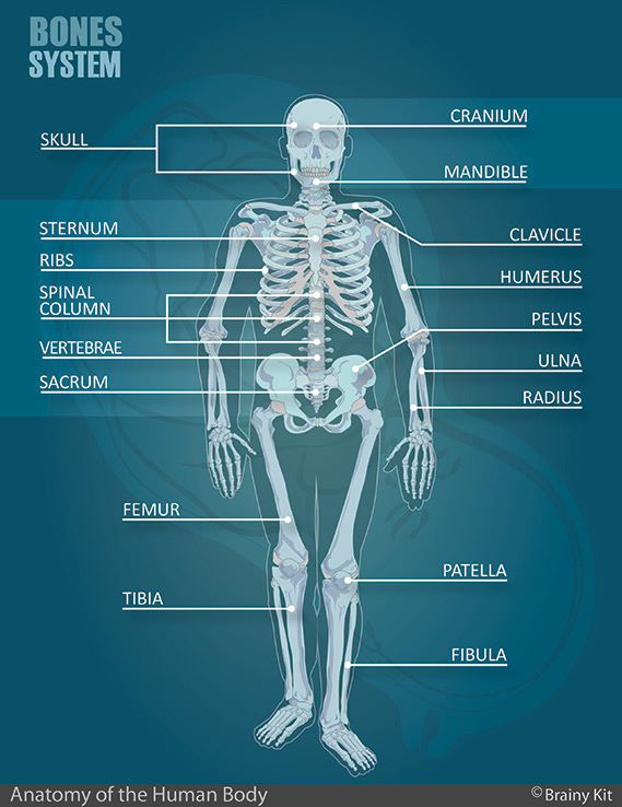
Anatomy For Kids Pdf Kit from getbrainybox.com Below is a quiz to test your knowledge on the human bones. This diagram depicts human bone anatomy. Some, like the rib cage, provide protection for softer body parts, while other bones enable mobility by supporting the muscles. As a nurse, you will need to know the basic about the human skeleton. In your anatomy & physiology lecture and lab class, you will be required to name each individual bone in the human body. There are 206 bones in the human skeleton, not including teeth and sesamoid bones (small bones found within cartilage): Added together, your bones make up about 15% of your body weight. The collection of bones in the human body is called the skeletal system.
The diaphysis and the epiphysis.the diaphysis is the tubular shaft that runs between the proximal and distal ends of the bone.
There also are bands of fibrous connective tissue —the ligaments and the tendons —in intimate relationship with the parts of the skeleton. The skeleton acts as a scaffold by providing support and protection for the soft tissues that make up the rest of the body. Human anatomy is the study of the shape and form of the human body. In this worksheet, we are going to review some of the major bones within the body. Human anatomy diagrams show internal organs, cells, systems, conditions, symptoms and sickness information and/or tips for healthy living. Über 7 millionen englischsprachige bücher. Human skeleton, the internal skeleton that serves as a framework for the body. It provides structure to the body, and each bone has a distinct purpose. The human body has four limbs (two arms and two legs), a head and a neck which connect to the torso. The structure of a long bone allows for the best visualization of all of the parts of a bone ().a long bone has two parts: Produces a collection of high quality casts of the human skeleton models for educators and practitioners in medicine, physical therapy, physical anthropology, comparative anatomy, and biomechanics. The vertebral column of the lower back includes the five lumbar vertebrae, the sacrum, and the coccyx. Human anatomy bone clones, inc.
Newborn babies are actually born with many more bones than this (around 300), but many bones grow together, or fuse, as babies become older. Human skeleton anatomy activity our bodies are more than they appear on the outside. The vertebral column of the lower back includes the five lumbar vertebrae, the sacrum, and the coccyx. 4.8 out of 5 stars. In this worksheet, we are going to review some of the major bones within the body.
3d Illustration Concept Of Human Skeleton System Appendicular Skeleton Anatomy Anterior View Canstock from comps.canstockphoto.com 2002, www.jurajartner.com page 15 atlas of human skeletal anatomy (arcus zygomaticus). It represents a vestigial tail, hence the common term tailbone. This framework consists of many individual bones and cartilages. It provides structure to the body, and each bone has a distinct purpose. The coccyx is a triangular arrangement of bone that makes up the very bottom portion of the spine below the sacrum. Its lower end helps create the knee joint. Altogether, the skeleton makes up about 20 percent of a person's body weight. Human skeleton anatomy activity our bodies are more than they appear on the outside.
Altogether, the skeleton makes up about 20 percent of a person's body weight.
Below is a quiz to test your knowledge on the human bones. Human anatomy bone clones, inc. There are 206 bones in the human skeleton, not including teeth and sesamoid bones (small bones found within cartilage): The skeleton acts as a scaffold by providing support and protection for the soft tissues that make up the rest of the body. Newborn babies are actually born with many more bones than this (around 300), but many bones grow together, or fuse, as babies become older. Human skeleton, the internal skeleton that serves as a framework for the body. The collection of bones in the human body is called the skeletal system. Human anatomy is the study of the shape and form of the human body. The patella, also called the knee cap, is a sesamoid bone. Added together, your bones make up about 15% of your body weight. Human skeleton anatomy activity our bodies are more than they appear on the outside. This includes the head, facial, hyoid, auditory, trunk, ribs, and sternum. Its lower end helps create the knee joint.
This framework consists of many individual bones and cartilages. Bones of the human skeletal system are categorized by their shape and function into five types. Did you know that they are made up of over 200 bones? 13 deep vein thrombosis varicose veins. Added together, your bones make up about 15% of your body weight.
Human Being Anatomy Skeleton Anterior View Image Visual Dictionary from www.ikonet.com Up bones has a broad coverage of the archaeological and anthropological aspects of excavating and interpreting human skeletal materials and is an excellent introductory text. In your anatomy & physiology lecture and lab class, you will be required to name each individual bone in the human body. 9 lobes of the brain. Bones of the pelvis and lower back the bones of the pelvis and lower back work together to support the body's weight, anchor the abdominal and hip muscles, and protect the delicate vital organs of the vertebral and abdominopelvic cavities. The coccyx is a triangular arrangement of bone that makes up the very bottom portion of the spine below the sacrum. Human anatomy diagrams show internal organs, cells, systems, conditions, symptoms and sickness information and/or tips for healthy living. This science quiz game will help you learn 15 of the most important bones. The femur, or thighbone, is the longest and largest bone in the human body.
Below is a quiz to test your knowledge on the human bones.
The patella, also called the knee cap, is a sesamoid bone. The femur is an example of a long bone. The body's shape is determined by a strong skeleton made of bone and cartilage, surrounded by fat, muscle, connective tissue, organs, and other structures. In your anatomy & physiology lecture and lab class, you will be required to name each individual bone in the human body. Bones of the human skeletal system are categorized by their shape and function into five types. 2002, www.jurajartner.com page 15 atlas of human skeletal anatomy (arcus zygomaticus). Altogether, the skeleton makes up about 20 percent of a person's body weight. The skeleton acts as a scaffold by providing support and protection for the soft tissues that make up the rest of the body. Human skeleton, the internal skeleton that serves as a framework for the body. It represents a vestigial tail, hence the common term tailbone. Individuals may have more or fewer bones than this owing to anatomical variations. The zygomatic bones form the alar ligaments (ligamenta alaria) together with the zygomatic process which connect the dens axis (second (processus zygomaticus) of each vertebra) to the occipital bone and temporal bone the zygomatic arch j.artner et al. There also are bands of fibrous connective tissue —the ligaments and the tendons —in intimate relationship with the parts of the skeleton.


0 Komentar